Skull Base Reconstruction
Endoscopic endonasal approaches are increasingly performed for the surgical treatment of multiple skull
base pathologies. Preventing postoperative CSF leaks remains a major challenge, particularly in extended approaches.
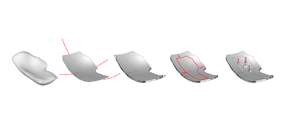
In this study, the authors assessed the potential use of modern multimaterial 3D printing and neuronavigation to help
model these extended defects and develop specifically tailored prostheses for reconstructive purposes.
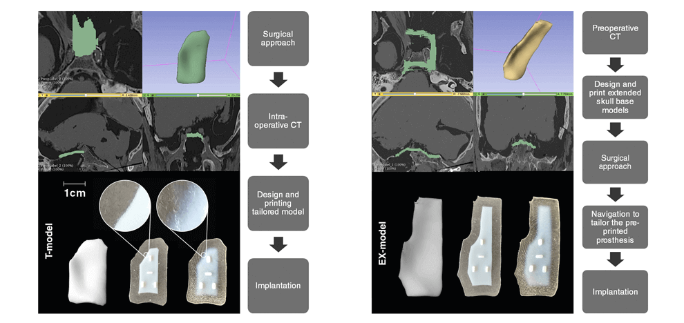
Extended endoscopic endonasal skull base approaches were performed on 3 human cadaveric heads. Preprocedure
and intraprocedure CT scans were completed and were used to segment and design extended and tailored
skull base models. Multimaterial models with different core/edge interfaces were 3D printed for implantation trials. A
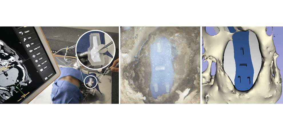
novel application of the intraoperative landmark acquisition method was used to transfer the navigation, helping to tailor
the extended models.
Prostheses were created based on preoperative and intraoperative CT scans. The navigation transfer offered
sufficiently accurate data to tailor the preprinted extended skull base defect prostheses. Successful implantation of the
skull base prostheses was achieved in all specimens. The progressive flexibility gradient of the models’ edges offered
the best compromise for easy intranasal maneuverability, anchoring, and structural stability. Prostheses printed based
on intraprocedure CT scans were accurate in shape but slightly undersized.
Preoperative 3D printing of patient-specific skull base models is achievable for extended endoscopic
endonasal surgery. The careful spatial modeling and the use of a flexibility gradient in the design helped achieve the
most stable reconstruction. Neuronavigation can help tailor preprinted prostheses.


